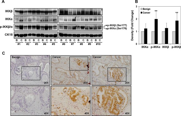
Figure 1. Expression of IKKα/β and their phosphorylation in various representative benign and prostate cancer tissues.

A. Protein expression of IKKα, IKKβ, p-IKKα (Ser176) and p-IKKβ (Ser177) in paired benign and cancer specimens was analyzed by Western blotting; cytokeratin18 expression served as loading control. A modest increase in IKKα and IKKβ expression was observed in cancer specimens compared to benign tissue; whereas a significant increase in p-IKKα and p-IKKβ was observed in cancer specimens. B. Relative density of bands showing protein expression in benign and cancer specimens. Mean ± SD; **P < 0.05, compared to benign tissue. C. Paraffin-embedded (4.0 μm) sections from benign and prostate cancer were used for p-IKKα/β (Ser177/176) expression by immunohistochemistry. Phosphorylated levels of IKKα/β was detected both in the nucleus and in the cytoplasm of malignant cells and was more intense in the cytoplasm. Magnified at x20 and x40. Details are described in ‘materials and methods’ section.
