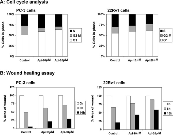
Figure 8. Effect of apigenin on DNA cell cycle and wound healing in prostate cancer cells.

A. DNA cell cycle analysis. PC-3 and 22Rv1 cells were synchronized in G0 phase by depleting the nutrients for 36 h (referred as control) and replating at sub confluent densities into complete medium containing vehicle or apigenin at indicated doses for 16 h, stained with PI (50 mg/ml) and analyzed by flow cytometry. Percentage of cells in G0-G1, S and G2-M phase were calculated using Mod-fit computer software and are represented in the right side of the histograms. A marked increase in G0-G1 phase accumulation of cells was observed after apigenin treatment. B. Wound healing assay. PC-3 and 22Rv1 cells were seeded into six-well plates and grown overnight. Then the cells were serum starved for 24 h. A sterile 200 μl pipette tip was used to scratch the cells to form a wound. The cells were washed with PBS and treated with vehicle or apigenin at indicated doses for 6 h and 16 h. Migration of the cells to the wound was visualized with an inverted Olympus phase-contrast microscope. A decrease in wound healing was observed after apigenin treatment in both cell lines. Details are described in ‘materials and methods’ section.
