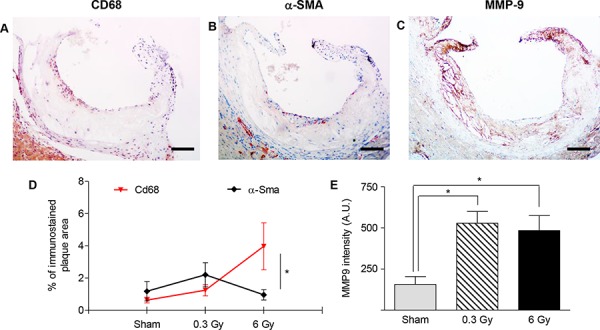
Figure 2. Plaques vulnerability after acute irradiation.

A–C. Representative sections of atherosclerotic plaques immunostained with antibodies against CD68 (A), α-SMA (B) and MMP-9 (C). Images refer to ApoE−/− mice 300 days after acute irradiation with 6 Gy. D. Mean percentage of total plaque area occupied by CD68- or α-SMA-positive cells. E. Intensity measurement of anti-MMP9 immunohistochemical staining by HistoQUEST software. Quantitative analysis involved 18 plaques/group. Data are shown as mean ± SEM. Differences were tested with Student's t-test. *P < 0.05. Bars: 100 μm.
