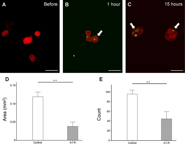
Figure 1. In vitro efficacy of S. typhimurium A1-R-GFP on HT-29-RFP colon cancer cells.

A–C. Confocal imaging of HT-29-RFP cells with S. typhimurium A1-R-GFP over time with the FV1000 confocal microscope. S. typhimurium A1-R infection of HT-29-RFP cells at one hour after administration (B) At 15 hour after administration, more bacterial cells were visualized in cancer cells (C). Arrows show infecting S. typhimurium A1-R expressing GFP. S. typhimurium A1-R inhibited cell proliferation both in colony area D. and number E. **P < 0.01. Error bars: ± 1 SE. Scale bars: 20 μm (BF, bright-field). The cells in Figures 1A, B and C were chosen as before and after examples of infection with S. typhimurium A1-R-GFP, not to indicate efficacy, which occurs at later times.
