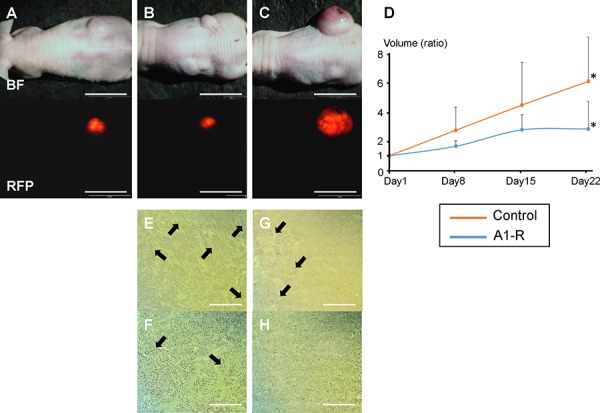
Figure 2. Efficacy of S. typhimurium A1-R on HT-29-RFP subcutaneous tumor growth.

Subcutaneous tumor models were established by injection of HT-29-RFP cells in the flanks of nude mice. A–C. Upper panels show bright-field images of tumor growth and lower show RFP images of tumor growth obtained with the OV-100 Small Animal Imaging System. An HT-29-RFP subcutaneous tumor in the right flank before treatment (day 1) (A), and after treatment with S. typhimurium A1-R at day 22 (B), HT-29-RFP tumor in the untreated control group at day-22 (C). D. S. typhimurium A1-R administration significantly decreased tumor volume at day-22 compared to no treatment. E–H. Representative histological images of excised tumors. S. typhimurium A1-R treated tumors had scattered necrosis surrounding viable cancer (E), (F). In contrast, untreated tumors had less necrosis (G), (H). (F) and (H) are high-magnification images of (E) and (G), respectively. *P < 0.05. Error bars: ± 1 SE. Arrows show necrotic areas. Scale bars: 20 mm (A) – (C), 500 μm (E) and (G), 200 μm (F) and (H).
