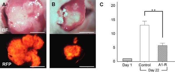
Figure 4. Efficacy of S. typhimurium A1-R on HT-29-RFP liver metastasis.

A, B. Upper panels are bright-field and lower panels are RFP images. Images of liver metastasis at day-22. No treatment (control group; A) and treated with S. typhimurium A1-R (S. typhimurium A1-R group; B). C. Bar graphs demonstrates the ratio of tumor fluorescent area at day 22 to day 1. Metastasis growth in the A1-R group was significantly inhibited compared to the untreated control group. **P < 0.01. Scale bars: 5 mm.
