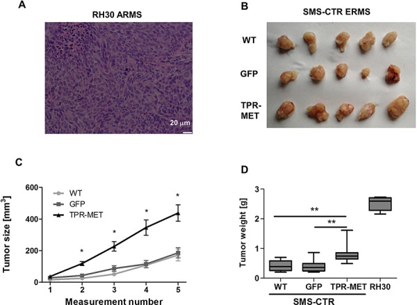
Figure 4. Activation of MET signaling in SMS-CTR ERMS cells enhances tumor growth in vivo.

A. Hematoxylin-eosin staining showed anaplastic morphology of tumors formed by RH30 ARMS cells with high basal MET level after subcutaneous implantation of the cells to immunodeficient NOD-SCID mice. B. Photograph of tumors formed by SMS-CTR cells isolated four weeks after subcutaneous implantation of the cells into NOD-SCID mice shows their differences in size. C. Tumor size was measured with caliper in different time points, n = 7–9. D. SMS-CTR and RH30 tumor weight was evaluated at the end of experiment, n = 4–9. Data in graphs are represented as mean +/− SEM.
