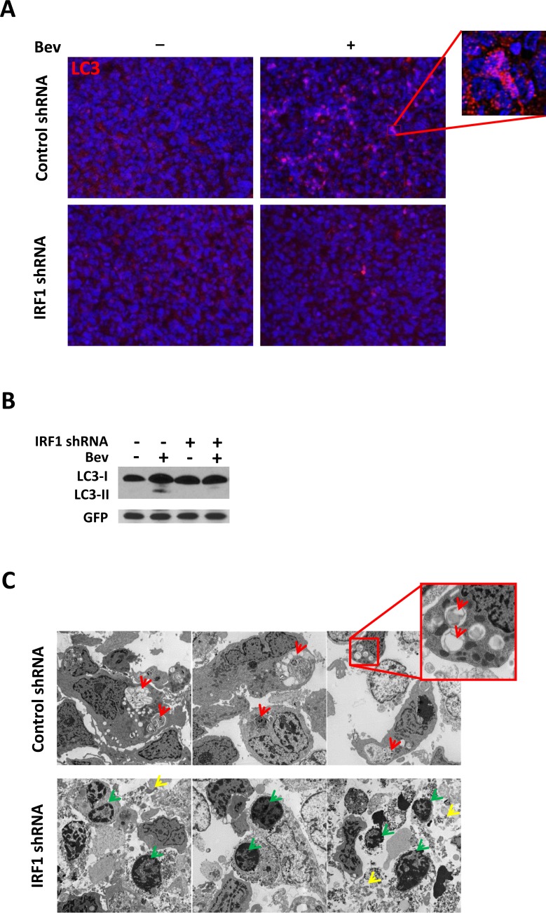
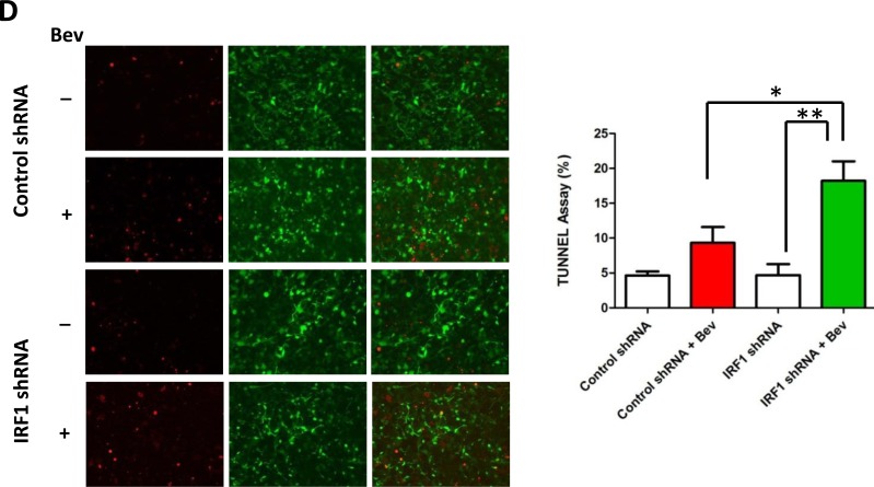
Figure 6. Inhibition of IRF1 leads to decreased autophagy and increased apoptosis in bevacizumab-treated gliomas.
A., Immunofluorescent staining of LC3 in glioma tumors with or without bevacizumab treatment. (at ×400 magnification) B., Western blot of LC3 conversion in bevacizumab-treated gliomas. Tissue proteins were collected from the tumor part of each brain. GFP-specific antibodies were used to assess protein loading for tumor cells. C., TEM analysis of autophagy and apoptosis in glioma tumors. Red arrows point to autophagic vacuoles while green arrows point to condensed chromatin at the margins of the nucleus, which is the hallmark of apoptosis. Loss of cellular integrity is indicated by yellow arrows. D., TUNEL assay measurement. Glioma tumors from each group were processed using a commercially available TUNEL assay kit. Red fluorescence indicates TUNEL-positive cell, green fluorescence indicates GFP-positive tumor cells. Both TUNEL and GPF positive cells were counted in nine different fields from three individual mice. *: P < 0.05, **: P < 0.01, P values were determined by Student's t test.


