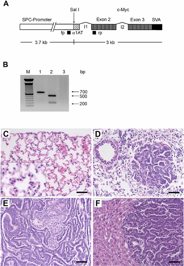
Figure 1. Histopathology of lung cancer and c-Myc transgene verification by PCR.

A. Scheme of the transgene for the production of transgenic mice. SPC promoter: human surfactant protein promoter; α1AT: first exon of the non-coding alpha 1 antitrypsin gene; I1: intron 1 of the alpha 1 antitrypsin gene fused to the first intron of the c-myc proto-oncogene; I2: intron 2 of the c-myc proto-oncogene; SVA: SV40 Poly A signal. The primer binding sites used for an identification of the transgene are indicated by black boxes: fp, forward primer; rp, reverse primer. B. Myc-transgene PCR of tail biopsy DNA. Lane 1–2: transgenic mice; lane 2: The amplified DNA was digested with Sal 1 to obtain fragments of about 200 and 500 bp; lane 3: non-transgenic controls; M, molecular weight standard. C. Normal subpleural parenchyma of non-transgenic controls. The insert represents a 2-fold magnification and depicted are normal pneumocytes with small regular nuclei. D. Illustrated is an initial papillary lung adenocarcinoma (PLAC) with real papilla, its own stroma and a size of 220 μm in diameter. Around the tumor numerous foci of dispersed AHH of the BAC-type are seen. E. Advanced PLAC with folded papillary structures of secondary and tertiary degree. F. Liver metastasis of PLAC. The bar represents 50 μm.
