
Figure 2. Hypoxia increases the percentage of CSCs in different breast cancer cell lines.
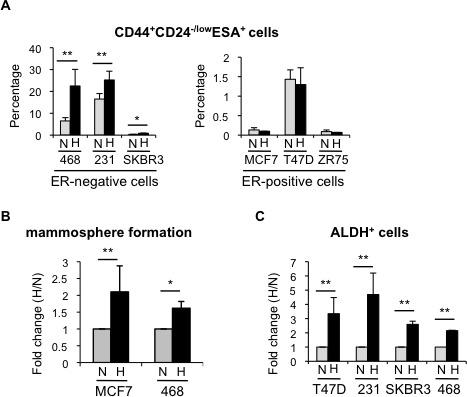
A. Percentage of CD44+CD24−/lowESA+ cells in ER-negative and ER-positive cell lines cultured in normoxia or hypoxia for 3 days. B. Number of mammospheres formed by MCF-7 or MDA-MB-468 cells cultured in normoxia or hypoxia and represented as fold change (hypoxia/normoxia). C. Percentage of ALDH+ cells in different cell lines cultured in normoxia or hypoxia. In A and B, means ±SD of at least three independent experiments are represented. *P < 0.05 **P < 0.01.