
Figure 3. Hypoxia reduces ER expression and transcriptional activity.
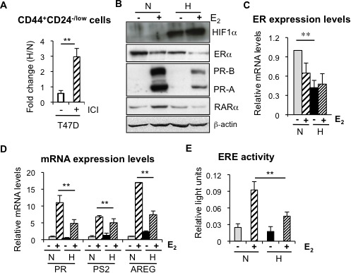
A. Percentage of CD44+CD24−/low cells in T47D cells treated or not with 0,5 μM fulvestrant (ICI 182,870) and cultured in normoxia or hypoxia. B. Representative western blot showing expression of ER and its targets PR and RARα in MCF-7 cells cultured under normoxic or hypoxic conditions, with or without 10 nM estrogen (E2). C. RNA expression levels of ER in MCF-7 cells treated or not with estrogen, in normoxia or hypoxia. D. RNA expression levels of PR, PS2 and AREG in MCF-7 cells treated or not with estrogen, in normoxia or hypoxia. In C, D, Data are presented as mean ±SD of 3 independent experiments. E. ER transcriptional activity in MCF-7 cells grown in normoxia or hypoxia in the presence of ethanol (−) or estrogen. The graph shows the mean ±SEM of 5 experiments done in triplicates. **P < 0.01.