
Figure 4. Hypoxia promotes dedifferentiation of breast cancer cells.
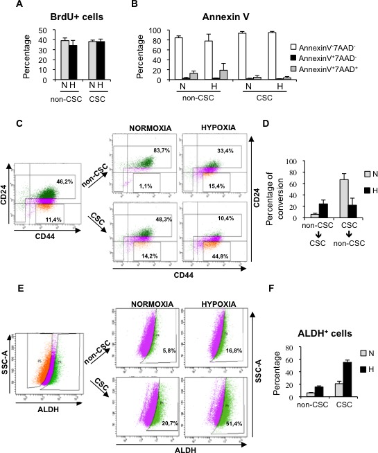
A. Percentage of BrdU positive MDA-MB-468 cells in sorted cell populations: CD44+CD24−/low CSCs and non-CSCs, grown in normoxia or hypoxia. B. Detection of apoptotic cells in CSCs and non-CSCs isolated from MDA-MB-468 cells that were grown in normoxia or hypoxia. The percentages of live cells (AnnexinV−7AAD−), early apoptotic (AnnexinV+7AAD−) and late apoptotic (AnnexinV+7AAD+) cells are shown. C. Representative example of a sorting experiment with CD44/CD24-stained MDA-MB-468 cells. On the left, CD44/CD24 staining of MDA-MB-468 cells cultured in hypoxia for 3 days. Gates used to sort CD44+CD24−/low cells (CSC) and the cell population depleted of CSCs (non-CSC) are presented. On the right, CD44/CD24 stainings performed with sorted CSCs and non-CSCs after 3 additional days growing in normoxia or hypoxia. D. Graph shows the capacity of CD44+CD24−/low CSCs to produce CD44+CD24high non-CSCs, and vice-versa, in normoxic and hypoxic conditions. E. Representative example of a dedifferentiation experiment performed with T47D cells sorted based on their ALDEFLUOR activity. F. Percentage of ALDH+ cells obtained after culturing ALDH+ CSCs and ALDH− non-CSCs in normoxia or hypoxia. A, B, D and F show means ±SD of three independent experiments.