
Figure 2. Treatment with triptolide selectively induces apoptosis of cultured and primary CD19+ CLL cells.
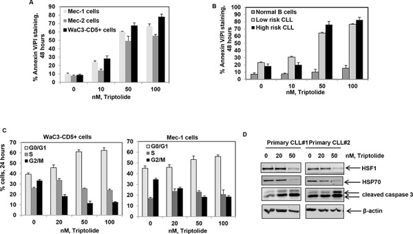
A. Percent apoptosis (Annexin V-FITC and PI staining) in Mec-1, Mec-2 and WAC3-CD5+ cells exposed to the indicated doses of triptolide for 48 hrs; bars indicate standard deviation. B. Percent apoptosis of CD19+ normal and primary CLL (both high and low risk) cells exposed to the indicated doses of triptolide for 48 hrs; bars indicate standard deviation. C. Percent of WaC3-CD5+ and Mec-1 cells in different phases of the cell cycle (G0/G1, S and G2/M) exposed to the indicated doses of triptolide for 24 hrs. D. Immunoblot analyses of HSF1, HSP70, cleaved caspase-3 and β-actin obtained from the cell lysates of CD19+ primary CLL cell samples treated with the indicated doses of triptolide for 24 hrs.