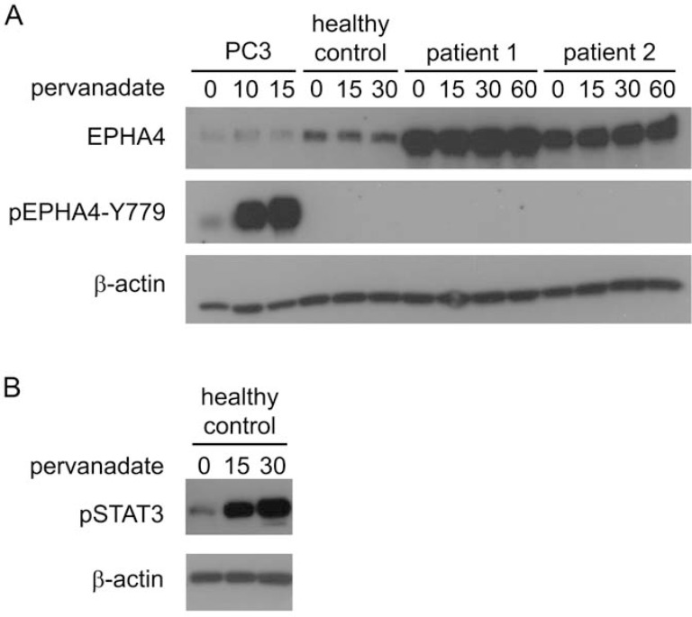
Figure 3. Activation of EPHA4 in CD4+ T-cells in vitro.
Cells were treated with pervanadate for the indicated time points (min) and were lysed for subsequent protein analysis with Western blot analysis. A. Expression of EPHA4 and phosphorylated EPHA4 (pEPHA4-Y779) in PC3 cells, and CD4+ T-cells isolated from peripheral blood from one healthy control and two SS patients. B. phosphorylated STAT3 (pSTAT3) in CD4+ T-cells isolated from peripheral blood from one healthy.

