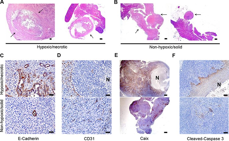
Figure 2. In vivo screening of PDAC cells with an in vivo preserved hypoxic/necrotic phenotype.

A–B. H&E–stained sections show centrally-necrotic tumors formed after orthotopic implantation of the hypoxic/necrotic cells and more solid forms of tumors formed by dedifferentiated cells, scale bar: 500 μm; arrow: tumor tissues; C. IHC with anti-E-Cadherin demonstrates a high proportion of anaplastic components in tumors developing from the hypoxic/necrotic cells, but not in tumors developing from the dedifferentiated cells, scale bar: 50 μm; D–F. IHC of anti-CD31 (scale bar: 50 μm), anti-Caix (scale bar: 500 μm) and anti-cleaved-caspase 3 (scale bar: 100 μm) show devascularized tumor regions, hypoxic zones and apoptosis in the necrotic tumors forming after transplantation of the hypoxic/necrotic cells, but not in the anaplastic tumors forming after transplantation of the dedifferentiated cells; N: necrosis.
