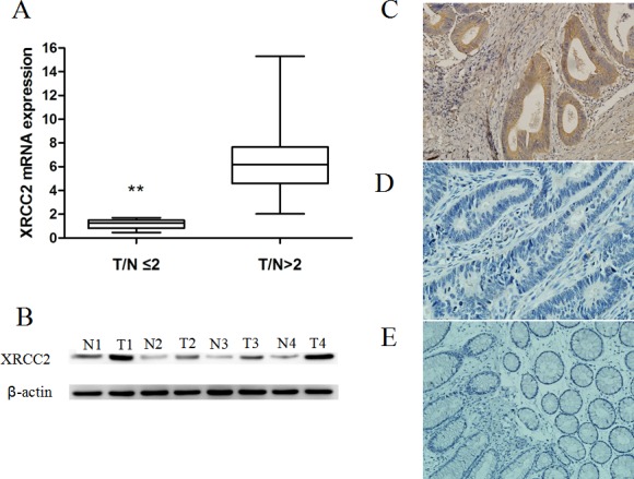
Figure 1. XRCC2 expression in the resected specimens that did not receive PRT.

A. Expression levels of XRCC2 mRNA were higher in the 50 rectal cancer samples compared with the 50 corresponding normal colorectal mucosa tissue samples (Wilcoxon signed rank test, **P < 0.01). B. Expression of XRCC2 was detected by Western blotting and higher levels were present in the rectal cancer samples (T1-T4) compared with the matched adjacent non-tumor tissues (N1-N4). C., D. Representative images of rectal cancer tissues that were positive C. and negative D. for XRCC2 expression in the immunohistochemical analysis performed (×200 magnification). E. A representative image of normal colorectal mucosa tissue that was negative for XRCC2 expression by immunohistochemistry (×200 magnification).
