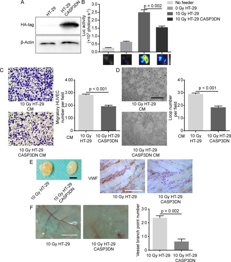
Figure 3. Caspase 3 in dying tumor cell mediates post-irradiation angiogenesis in vitro and in vivo.
A. Dominant-negative caspase 3 expression was confirmed by Western blot analysis (HA-tag was fused with dominant-negative caspase 3 in tandem). B. Proliferation-stimulating effect of dying HT-29 CASP3DN cells was highly inferior to that of dying parental HT-29 cells. C. Left panel, representative images of HUVEC migration to different indicated CM. Scale bar: 250 μm. Right panel, quantification of HUVEC migration to indicated CM. D. Left panel, representative images of tube formation of HUVECs in different indicated CM. Scale bar: 250 μm. Right panel, loop number quantification of tube formation assay performed in different CM. E. Matrigel mixed with 10 Gy-irradiated HT-29 cells or HT-29 CASP3DN cells was subcutaneously injected into flank of nude mice for 8 days. Left panel, representative images of plugs mixed with indicated cells. Scale bar: 5 mm. Right panel, immunohistochemical analysis of plug sections for VWF staining. Scale bar: 125 μm. F. Left panel, representative images of skin vasculature adjacent to indicated plugs. Scale bar: 5 mm. Right panel, quantification of dermal blood vessel adjoining indicated plugs.

