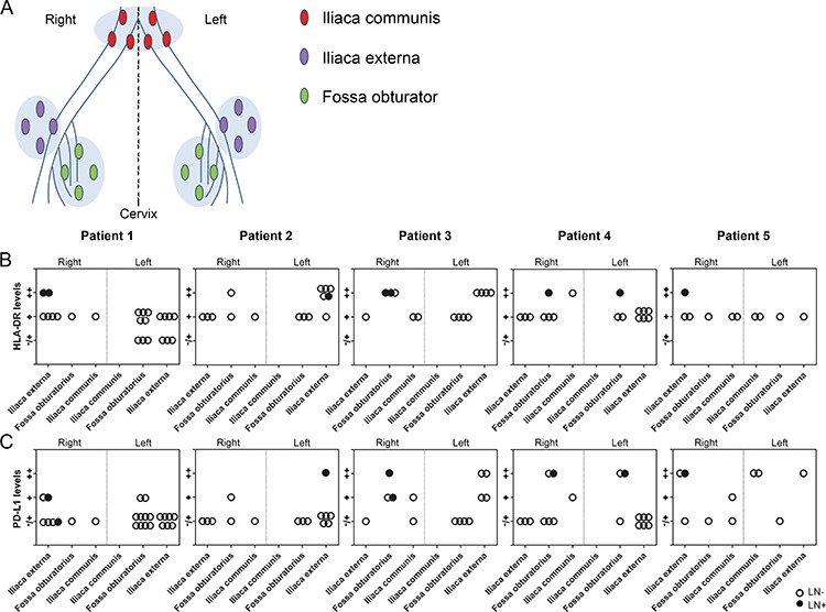
Figure 4. HLA-DR and PD-L1 levels in the lymphatic basins of five patients with cervical cancer.

A. Reconstruction of the anatomical locations of pelvic lymph nodes from five patients with cervical cancer, showing the following regions: iliaca externa (purple), fossa obturator (green) and iliaca communis (red) on both sides (right and left). Graphs showing B. HLA-DR levels and C. PD-L1 levels (in paracortical areas in tumor-negative lymph nodes (LN−) and in case of tumor-positive lymph nodes (LN+), paracortical and peritumoral areas) per lymphatic basin per patient. Levels for HLA-DR and PD-L1 are indicated by (−/+) minimal, (+) moderate, and (++) high numbers of positive cells. Closed circles represent LN+, open circles represent LN−.
