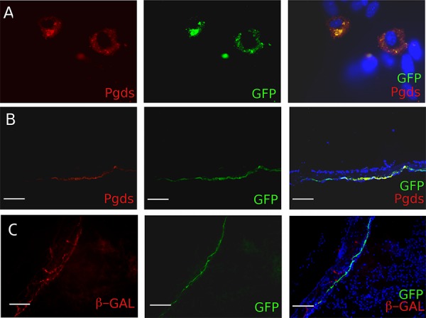
Figure 2. In vitro and in vivo characterization of the PDGDStv-a mouse.

A. Functional analyses of the PGDStv-a transgenic construct in a culture of arachnoidal cells. Infection with RCAS-eGFP was directed to cells with arachnoid morphology and the enhanced green fluorescent protein (eGFP)-infected cells co-expressed PGDS (red cytoplasmic staining). B. In vivo characterization of PN7 PGDStv-a mouse brains and meninges. Arachnoid PGDS positive cells forming a thin layer were found to be positive for GFP after transfection with RCAS-eGFP. C. Co-localisation of X-gal (red staining) and GFP stainings in PGDS positive arachnoid cells demonstrating the possibility to co-transfect the same cells with both RCAS and Adenovirus vectors.
