
Figure 1. LYAR is highly expressed in human metastatic CRC tissues.
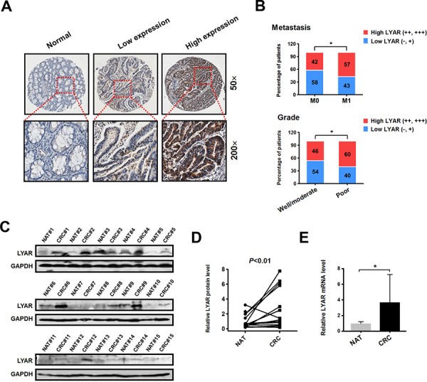
A. Immunohistochemical staining of the LYAR protein in adjacent normal and carcinoma tissues from colorectal cancer patients. Representative micrographs are shown at the original magnification (50 × and 200 × ) as indicated. Low (−, +) and high LYAR expression (++, +++) were classified according to the LYAR immunostaining scores, IRS. B. The percentage of patients with different metastasis statuses (M0, no regional or distant metastasis; M1, regional or distant metastasis) and differentiation grades as scored by IRS. *P < 0.05 compared with the indicated group. C. Western blot analysis of LYAR in cell lysates from normal adjacent tissues (NAT) and colorectal tumor tissues (CRC) (n = 15). GAPDH served as a loading control. D. Quantitation of the density of the protein bands from the western blots in (C); P < 0.01 compared with the paired NAT. E. Quantitation of the LYAR mRNA levels normalized to GAPDH mRNA levels from the tissues from in (C). *P < 0.05 compared with the NAT control.