
Figure 4. OVA intestinal allergy promote colorectal carcinogenesis in HDC−/− mice.
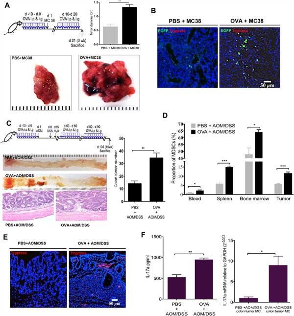
A. Tumor diameter from MC38 colon carcinoma tumor cells injected HDC−/−;HDC-GFP mice. Mice (n = 8/group) treated with OVA exhibited bigger tumor diameter than PBS controls. B. Increased accumulation of MCs and HDC-EGFP+ myeloid cells within MC38 carcinoma in the group of mice treated with OVA. C. Schematic representation of the OVA immunization plus AOM/DSS carcinogenesis protocol (Upper panel). Representative macroscopic pictures and H&E staining images from paraffin embedded colon tissue of tumor mice (n = 10/group, Lower panel). The number of tumors with statistics presented by bar picture (Right). D. Flow cytometry analysis of CD11b+Gr1+ MDSCs proportion in CD45+ cells in OVA or PBS plus AOM/DSS treated mice (n = 10/group) in blood, spleen, bone marrow, and colon tumor. E. Representative tryptase immunofluorescence staining from tumor-bearing mice colon frozen sections. F. OVA immunization increases serum IL-17a level and IL-17a mRNA expression in sorted tumor MCs from AOM/DSS colorectal carcinogenesis mice (n = 10/group). Data are representative from two independent experiments.