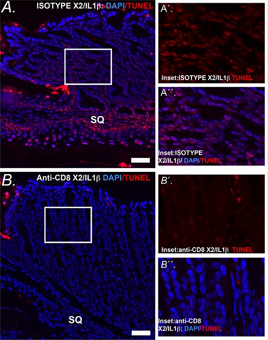
Figure 13. Apoptosis at the SCJ in K14-Cdx2::L2-IL-1β mice is lost with knockdown of CD8+ cells.

A. Representative image of combined nuclear (DAPI-blue) and TUNEL (Tetramethylrhodamine –red) at the SCJ of isotype treated K14-Cdx2::L2-IL-1β mice. (x200, white bar = 50 μm) SQ = squamous forestomach. A′ inset. Representative higher-power imaging for TUNEL staining of the apoptotic columnar metaplasia at the SCJ. A′′ inset. similar higher-power imaging for nuclei (DAPI-blue) and TUNEL (Tetramethylrhodamine –red). There is a highly significant TUNEL labeling of the nuclei of the glandular epithelial cells of isotype treated K14-Cdx2::L2-IL-1β mice n = 3 K14-Cdx2::L2-IL-1β mice for each treatment. B. Representative imaging of combined nuclear (DAPI-blue) and TUNEL (Tetramethylrhodamine –red) of the SCJ sections from a mouse treated with the anti-CD8 antibody demonstrating significantly reduced TUNEL+ nuclei in both the squamous and glandular compartments. (x200, white bar = 50 μm) SQ = squamous forestomach. B′ inset. Representative higher-power imaging demonstrating little TUNEL staining of the columnar metaplasia at the SCJ. B′′ inset. similar higher-power imaging for nuclei (DAPI-blue) and TUNEL (Tetramethylrhodamine –red) revealing little TUNEL labeling of the nuclei of anti-CD8 antibody treated K14-Cdx2::L2-IL-1β mice n = 3 K14-Cdx2::L2-IL-1β mice for each treatment.
