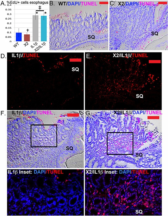
Figure 6. Apoptosis but not cell proliferation is significantly altered in the SCJ metaplasia K14-Cdx2::L2-IL-1β transgenic mice.

A. Average counts of EdU incorporation in the esophagi of transgenic and control mice. EdU was detected by Alexa Fluor 594 azide (red) and cell nuclei were stained with DAPI (blue). At least 300 cells per mouse were counted. L2-IL-1β over-expression promotes cell proliferation in esophageal epithelium. * Significantly differs from control WT mice by one-sided ANOVA and Tukey's Multiple comparison test adjusted p < 0.043. ‡ no significant difference between double and single L2-IL-1β TG mice adjusted p = 0.9996; † no significant difference between K14-Cdx2 and WT mice, adjusted p = 0.9932. n = 15 mice WT, n = 8 mice K14-Cdx2, n = 15 mice L2-IL-1β, and n = 10 K14-Cdx2::L2-IL-1β mice. B. and C. Representative imaging of combined nuclear (DAPI-blue), TUNEL (Tetramethylrhodamine –red) and differential interference contrast microscopy (Nomarski) of the SCJ of (B) WT and (C) K14-Cdx2 mice. (x200, red bar = 50 μm). D. and E. Representative imaging TUNEL (Tetramethylrhodamine –red) staining of the SCJ metaplasia of (D) L2-IL-1β and (E) K14-Cdx2::L2-IL-1β mice. There is a highly significant increase in TUNEL labeling of the nuclei of the glandular epithelial cells. (x200, red bar = 50 μm). F. and G. Representative imaging of combined nuclear (DAPI-blue), TUNEL (Tetramethylrhodamine –red) and differential interference contrast microscopy (Nomarski) of the SCJ metaplasia of (F) IL-1β and (G) K14-Cdx2::L2-IL-1β mice. (×200, red bar = 50 μm). Inset: ×400 magnification of SCJ metaplasia with only nuclear (DAPI-blue) and TUNEL (Tetramethylrhodamine –red) staining illustrate the localization of TUNEL staining to nuclei, including the glandular epithelium of the metaplasia.
