
Figure 5. IKKβ expression in microglia/macrophages in low and high grade gliomas.
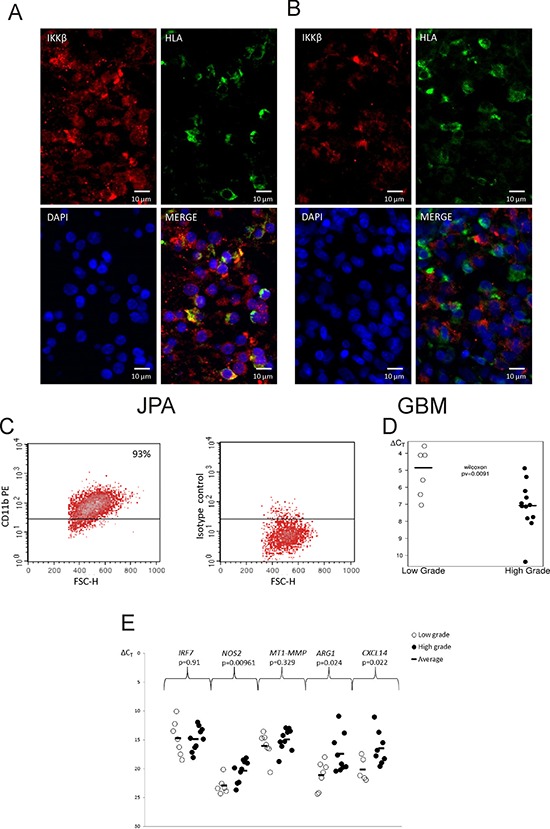
A, B. Images of double staining for CD11b+ (red) and IKKβ (brown) performed on sections of GBM (A) and JPA (B); scale bars 100 μm (insets: 50 μm). C. Microglia/macrophages were separated from freshly resected tumors using a magnetic-bead-conjugated anti-CD11b antibody. Purity of the positive fraction stained with anti-CD11b-PE antibody after magnetic separation was 93%. D–E. Quantification of the IKBKB (D) and M1/M2 marker gene expression (E) in CD11b+ cells isolated from low and high grade gliomas. Lower ΔCT is consistent with higher gene expression. Statistical analysis was done with the Wilcoxon testand t test, respectively.