
Figure 5. Effects of PIM kinase inhibition on primary leukemic progenitors from CML patients.
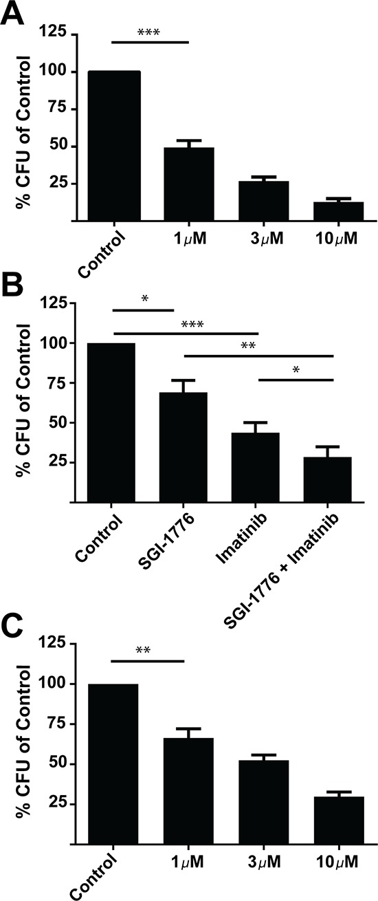
A. Primary mononuclear cells derived from CML patient samples were plated in methylcellulose in the presence of control (DMSO) or SGI-1776 at the indicated concentrations for approximately fourteen days. Leukemic progenitor colony formation (CFU-GM) was assessed in clonogenic assays in methylcellulose. Data are expressed as a percentage of DMSO control-treated cells. Shown are the means + SE of five experiments performed with samples from five different patients. ***p < 0.001 using a paired t-test. B. Primary mononuclear cells derived from CML patient samples were plated in methylcellulose in presence of control (DMSO), SGI-1776 (0.5 μmol/L) or imatinib (0.5 μmol/L) alone or in combination for approximately fourteen days, as indicated. Leukemic progenitor colony formation (CFU-GM) was assessed in clonogenic assays in methylcellulose. Data are expressed as a percentage of DMSO control-treated cells. Shown are the means + SE of five experiments performed with samples from five different patients. *p < 0.05, **p < 0.01, ***p < 0.001 using a paired t-test C. Primary mononuclear cells derived from CML patient samples were plated in methylcellulose in the presence of control (DMSO) or AZD-1208 at the indicated doses for approximately fourteen days. Leukemic progenitor colony formation (CFU-GM) was assessed in clonogenic assays in methylcellulose. Data are expressed as a percentage of DMSO control-treated cells. Shown are the means + SE of four experiments performed with samples from four different patients. **p < 0.01 using a paired t-test.