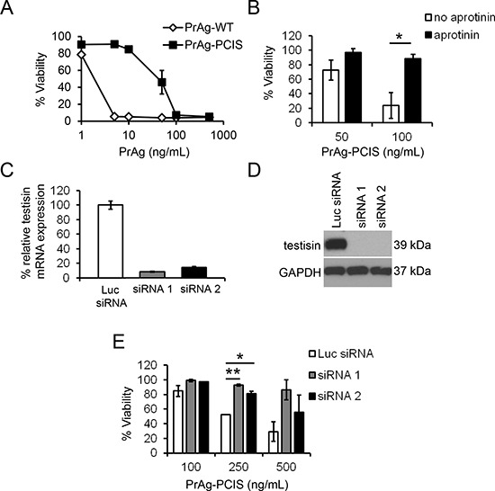
Figure 5. Endogenous testisin activity activates the PrAg-PCIS toxin and promotes HeLa tumor cell killing.

A. HeLa cells are sensitive to the PrAg-PCIS toxin. HeLa cells were incubated with 0–500 ng/mL of PrAg proteins (PrAg-PCIS or PrAg-WT) and FP59 (50 ng/mL) for 48 hours and then assayed for cell viability by MTT assay. Values are calculated from two independent experiments performed in triplicate B. Aprotinin-sensitive proteases contribute to PrAg-PCIS toxin-induced cytotoxicity. HeLa cells were pre-incubated in the presence of a final concentration of 100 μM aprotinin for 30 minutes, prior to treatment with the indicated concentrations of PrAg-PCIS and FP59 (50 ng/mL) for 2 hours. Media was replaced and cell viability assayed 48 hours later by MTT assay. Values are calculated from two independent experiments performed in triplicate. *p < 0.05. C. siRNA knockdown of testisin mRNA expression in HeLa cells. mRNA expression levels are normalized to GAPDH and expressed relative to the Luc-siRNA control. D. Immunoblot analysis of testisin protein expression after siRNA knockdown. The blot was probed using anti-testisin antibody and reprobed with anti-GAPDH antibody. Data is representative of at least two independent experiments. E. Depletion of testisin reduces the sensitivity of HeLa cells to PrAg-PCIS toxin-induced cytotoxicity. Testisin siRNA or control Luc-siRNA transfected HeLa cells were incubated for 6 hours with indicated concentrations of PrAg-PCIS and FP59 (50 ng/mL). Media was replaced and cell viability was assayed 48 hours later by MTT assay. Values are the means calculated from two independent experiments performed in triplicate. *p < 0.05; **p < 0.01.
