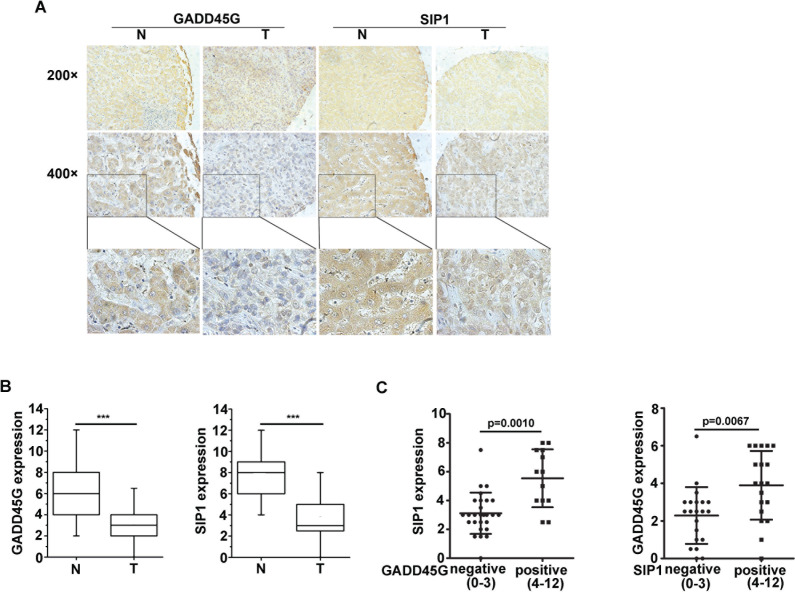
Figure 6. Expression levels of GADD45G and SIP1 were coincidently downregulated in human hepatocellular carcinoma.
(A) Representative images of GADD45G and SIP1 immunohistochemical staining. Original magnification, ×200 (upper panel); ×400 (middle panel). (B) Box-and-whisker plots of the staining of GADD45G and SIP1. The immunoreactive score is shown as mean ± SD (***P < 0.001). SD, standard deviation. (C) Statistical analysis of SIP1 expression in HCC sections negative (score, 0-3) and positive (score, 4-12) for GADD45G staining (left panel; P = 0.0010). SIP1-negative HCC sections exhibited a significantly lower GADD45G staining (right panel; P = 0.0067). The immunoreactive score is shown as mean ±SD. SD, standard deviation.

