
Figure 2. TAM-derived HO-1 modulates prostate cancer progression.
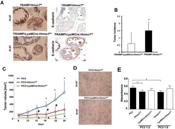
A–B. Hmox-1fl/fl mice were crossed with LyzM-Cre mice to generate myeloid linage specific knockout of HO-1 (LyzM-Cre:Hmox1fl/fl), which were further crossed to TRAMP mice. Tumor initiation was measured at 25 weeks of age by evaluation of Ki67 positivity within the prostate glands. Sections were also stained with E-cadherin, loss of which is a marker of tumor progression. Number of glands per field of view (FOV) with prostatic intraepithelial neoplasia (PIN) or adenocarcinoma lesions were evaluated as incidence of tumorigenesis based on H&E staining in n = 5–6 mice per group. Data ± SD are shown, *p < 0.05 (one-tailed t-test). C–D. PC3 cells or PC3 cells mixed with BMDM from Hmox1fl/fl and LyzM-Cre:Hmox1fl/fl mice were inoculated into the flanks of nude mice. Xenograft growth was measured over time (C) and Ki67 staining (D) was performed at day 26 after inoculation. *p < 0.01, PC3 versus PC3+ BMDM from Hmox1fl/fl mice and #p < 0.05, PC3+ BMDM from Hmox1fl/fl versus PC3+ BMDM from LyzM-Cre:Hmox1fl/fl; n = 3–5 mice per group. E. BrdU incorporation assay was employed to measure proliferation of PC3 co-cultured with BMDM from LyzM-Cre:Hmox1fl/fl and Hmoxfl/fl mice in the ratio of 1:2 or 1:5 (PC3:BMDM) for 24 h. Data are representative for 3 independent experiments in triplicates. *p < 0.05.