
Figure 3. Presence of HO-1-postitive macrophages in the tumor microenvironment modulates infiltration of Gr1+ and NK cells.
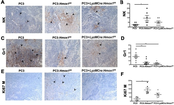
A–F. Immunohistochemistry of xenografts established from PC3 cells alone or mixed with BMDM (1:1) from Hmox1fl/fl and LyzM-Cre:Hmox1fl/fl mice in nude mice as in Figure 2. NK cells (NKp46, A–B), granulocyte (Gr-1, C–D) or proliferation (mouse Ki67) (E–F) markers were evaluated by immunohistochemistry. Quantization of staining is expressed as number of positive cells (Ki67, Gr1) or % of area of staining (NKp46, Gr1) presented from n = 5–6 FOV (B, D, F). n = 3–4 animals per group. Data are presented as mean values ±SD.