
Figure 4. AR-v7 ubiquitination results in AR-v7 localization to cytoplasm for protein degradation.
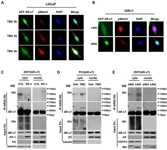
A. LNCaP cells were transfected with GFP-tagged AR-v7 plasmid and then treated with 5μM of TMC for 0, 2 or 4 hours. B. 22Rv1 cells were transfected with GFP-tagged AR-v7 plasmid together with either cAkt or dAkt expression vector for 24 hours. Cells were fixed, immunostained with phosphor-Mdm2(ser166) antibody, and examined by fluorescence microscope. 293T cells were transfected with AR-v7 plasmid (C and E). PC3 cells were stably introduced with exogenous AR-v7 protein D.. Cells were co-transfected with either control or PP-1 plasmid C., or treated with vehicle or 5μM of TMC D. or transfected with dAkt or cAkt expression vector for 24 hours E.. Cells were also treated with 2μg/ml of MG132 for another 16 hours. Cytoplasmic and nuclear fractions of protein lysis were extracted. Histone 3 (H3) and tubulin were detected by immunoblotting and were used as markers to confirm the efficacy of cytosol and nuclear fraction. In vivo ubiquitination assays were performed as described in the Material and Method section. All experiments were repeated at least three times with one set of results shown in the figure.