
Figure 1. Comparative analysis of Tim-3 expression and surface presence in primary human AML cells and healthy leukocytes.
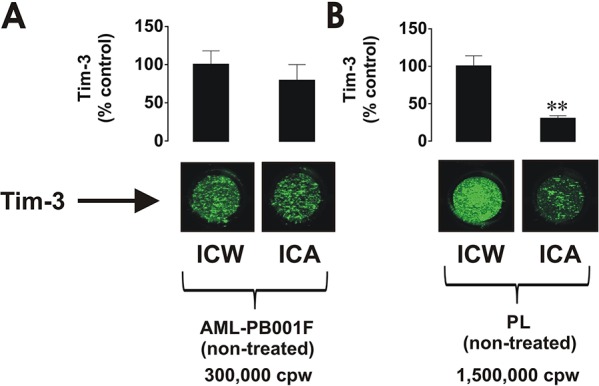
300,000 per well of AML-PB001F primary human AML cells A. and 1,500,000 per well of healthy PLs B. were subjected to in-cell Western (ICW) in order to detect total Tim-3 expression. The Tim-3 surface presence was analysed by in-cell assay (ICA). For healthy PLs, 1,500,000 cells per well were necessary to properly visualise Tim-3 receptor on the cell surface. Fluorescence values obtained for AML cells and healthy PLs were divided by respective cell number (300,000 or 1,500,000) and used for calculations. Images are from one experiment representative of three which gave similar results. Quantitative data are shown as means ± SEM of at least three individual experiments; **p < 0.01 vs. control.