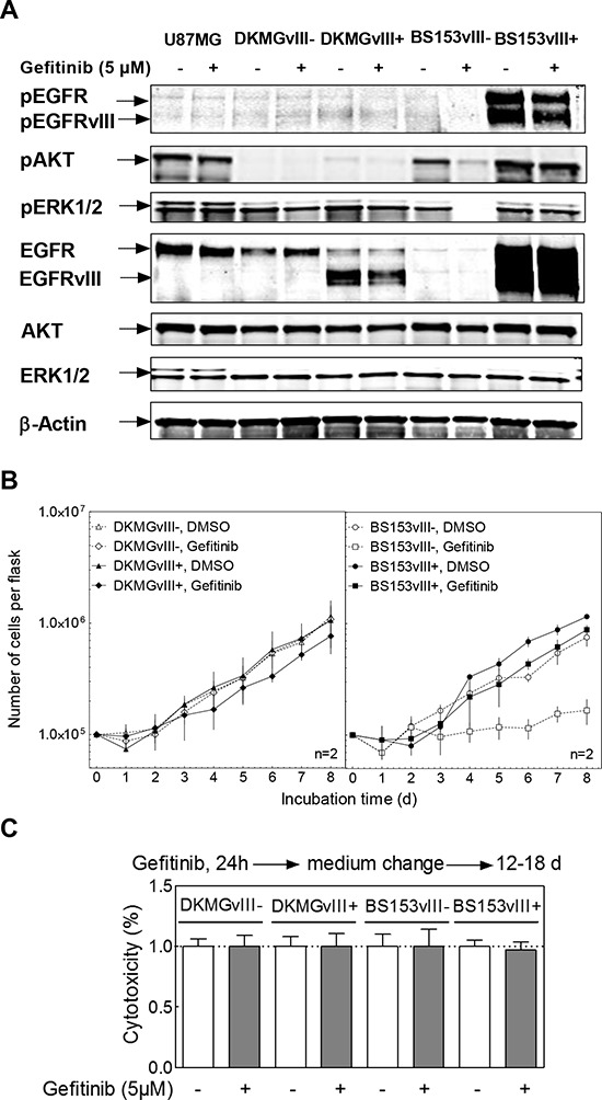
Figure 5. Effect of gefitinib on EGFR signaling, proliferation and clonogenicity.

DKMGvIII−/+ and BS153vIII−/+ cells were treated with 5 μM gefitinib. A. After 2 h incubation, phosphorylation of EGFR (Y1173), AKT (T308) and ERK1/2 (T202/Y204) was determined by Western blot analysis using phosphospecific antibodies. The detection of unphosphorylated proteins and actin served as controls. B. Proliferation of DKMGvIII−/+ and BS153vIII−/+ cells in the presence of gefitinib (n = 2). The cell number was determind for up to 8 days. The data set from Figure 3C was used for comparison with untreated cells. C. Relative cytotoxicity of gefitinib as determind by colony forming assay (pre-plating). The surviving fraction of gefitinib-treated cells was normalized to the plating efficiency of untreated cells.
