
Figure 4. The binding of anti-CD146 mAb (A) and anti-CD146 coated nanoparticles (B) in vivo was studied using fluorescence microscopy.
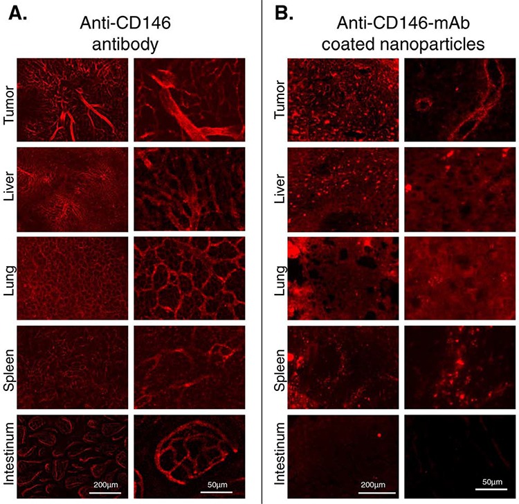
Injection of mAb resulted in the staining of blood vessels in tumor, liver, lung, spleen and intestinal mucosa (A) Anti-CD146 coated nanoparticles were distinctly bound to tumor endothelium, whereas low binding in peritumoral liver was found (B) Images of two different magnifications are shown (see respective vertical rows) and magnification bars (50 or 200 μm) are indicated.