
Figure 4. Induction of IL-6 secretion by TrkB enhances nuclear translocation of STAT3 and upregulates Twist-1 and Twist-2.
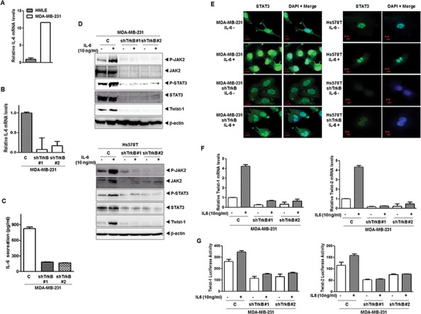
A. The relative expression of mRNA encoding IL-6 in MDA-MB-231 cells relative to that of HMLE cells, as determined by quantitative RT-PCR. The 18S mRNA level was used to normalize the variability in template loading. B. The relative expression of the mRNA encoding IL-6 in MDA-MB-231 control-shRNA or TrkB-shRNA cells, as determined by quantitative RT-PCR. The 18S mRNA level was used to normalize the variability in template loading. C. IL-6 secretion by MDA-MB-231 control-shRNA or TrkB-shRNA cells, as determined by ELISA. The data are reported as the means ± SEM. D. Western blot analysis of the expression of phospho-JAK2, JAK2, phospho-STAT3, STAT3, and Twist-1 proteins in MDA-MB-231 and Hs578T control-shRNA or TrkB-shRNA cells treated with or without IL-6. β-actin was used as a loading control. E. Immunofluorescence images of STAT3 in MDA-MB-231 and Hs578T control-shRNA or TrkB-shRNA cells treated with IL-6. The green signal represents staining of the corresponding protein, while the blue signal represents DAPI staining. F. The relative expression levels of mRNA encoding Twist-1 and Twist-2 in MDA-MB-231 control-shRNA or TrkB-shRNA cells treated with IL-6, as determined by quantitative RT-PCR. The 18S mRNA level was used to normalize the variability in template loading. G. Promoter activity of Twist-1 and Twist-2 genes in MDA-MB-231 control-shRNA or TrkB-shRNA cells treated with or without IL-6. Each bar represents the mean ± SEM of three experiments.