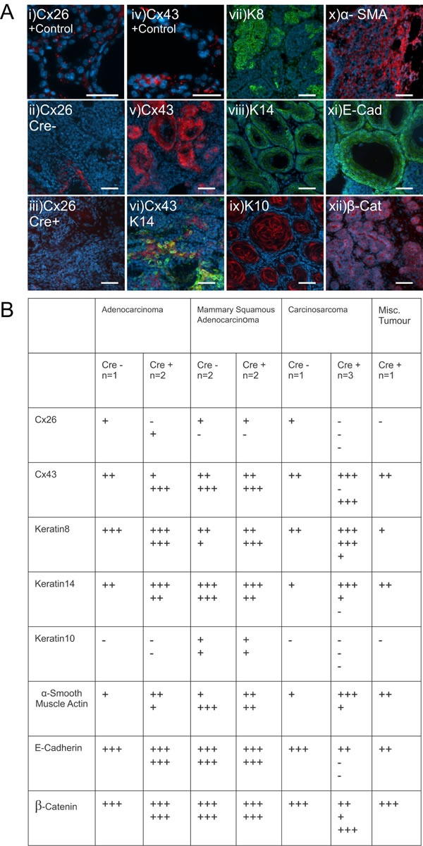
Figure 5. Mammary tumors from Cx26 knockout mice express similar epithelial protein markers as control mice.

A. Representative images of paraffin-embedded primary mammary tumor sections immunolabelled with luminal and myoepithelial markers that included; Cx26 (i, ii, iii, red), Cx43 (iv, v, vi, red), keratin 14 (vi, viii green), keratin 8 (vii, green), keratin 10 (ix, red), α-smooth muscle actin (x, red), E-cadherin (xi, green) and β-catenin (xii, red). Hoechst denotes nuclei. Scale bars=50 μm. B. Table indicates relative number of cells that are positive for the luminal and myoepithelial markers based on immunofluorescent labelling. +++50-100%, ++11-49%, +1-10%, −0%.
