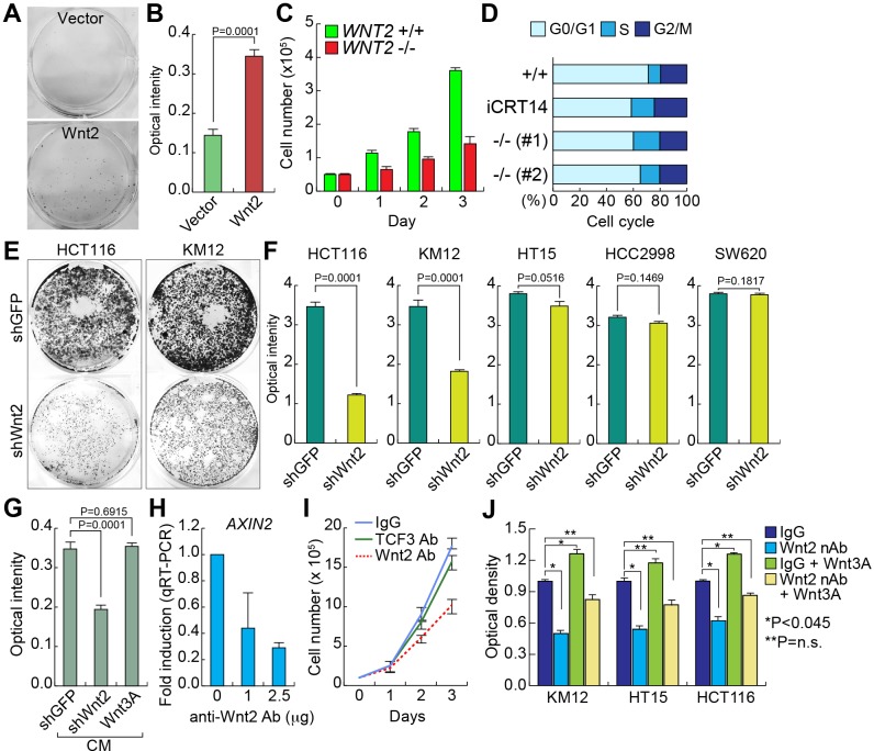
Figure 3. Wnt2 is required for CRC cell proliferation.
A. and B. Increased cell proliferation by Wnt2. CCD841CoN IECs were stably transfected with Wnt2-expressing plasmid. Then, the equal number of cells were plated and cultured for 20 days. Crystal violet staining A.; quantification of optical intensity of crystal violet stained cells B.. C. Decreased cell proliferation by WNT2 KO. The equal number of HCT116 (WNT2 WT vs. WNT2 KO) cells were plated and counted in a different time course. D. Cell cycle analysis of WNT2 KO cells. HCT116 cells were analyzed using flow cytometry. E. and F. Depletion of Wnt2 reduced CRC cell proliferation. CRC cells were stably transduced with lentivirus encoding shRNAs (shGFP: control; shWnt2). Then, the equal number of cells were plated and grown for 14 days for crystal violet staining E. and quantification F.. G. Decreased cell proliferation by Wnt2 KD CM. KM12 cells were transfected with plasmids (empty vector: control; shWnt2; Wnt3A). After transfection, KM12 cells were incubated with serum free DMEM for collection of CM. Then, each CM was treated to HCT116 cells. Cell proliferation (crystal violet staining quantification) was monitored after were monitored for 20 days of CM treatment. H. Downregulation of AXIN2 by neutralizing Wnt2 protein. HCT116 cells were treated with anti-Wnt2 antibody. 24 hours after treatment, cells were analyzed for qRT-PCR of AXIN2. HPRT served as an internal control. I. Inhibition of cell proliferation by neutralization of Wnt2. The equal number of HCT116 cells were plated and treated with antibodies (IgG and TCF3 antibodies: negative controls; Wnt2 antibody). Cell proliferation was analyzed by cell counting. J. Rescue of Wnt nAb-induced cell growth inhibition by Wnt3A. CRC cells were treated with Wnt2 nAb and/or Wnt3A (100 ng/ml). Cell proliferation was analyzed by crystal violet staining and quantification using plate reader.

