
Figure 3. CL4 hampers EGFRvIII activation.
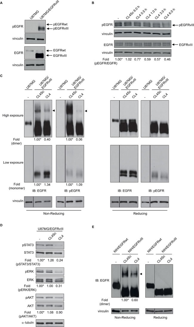
Lysates from A. U87MG and U87MG/EGFRvIII cells maintained in 2% FBS-containing medium for 6 hours or from B. U87MG/EGFRvIII cells maintained in 2% FBS-containing medium for 6 hours and then left untreated for further 6 hours or treated with 200 nmol/l CL4 or CL4Sc for the indicated times, were immunoblotted with anti-pEGFR and anti-EGFR antibodies, as indicated. In B, values below the blot indicate the ratio of pEGFR to total EGFR signal levels, normalized to the respective vinculin signal level, and reported as relative to untreated cells, arbitrarily set to 1 (labeled with asterisk). C. Equal amounts of lysates from U87MG or U87MG/EGFRvIII cells, maintained in 2% FBS for 6 hours and then treated with 200 nmol/l CL4 or CL4Sc for 24 hours, were run on 6% SDS-PAGE under non-reducing (left) and reducing (right) conditions and immunoblotted with anti-EGFR and anti-pEGFR antibodies. D. Lysates from U87MG/EGFRvIII cells treated as in “C” were immunoblotted with anti-pSTAT3, anti-pERK and anti-pAKT antibodies, as indicated. Filters were stripped and reprobed with anti-STAT3, anti-ERK and anti-AKT antibodies. E. Lysates from NIH/EGFRwt or NIH/EGFRvIII cells treated as in “C” were immunoblotted with anti-EGFR antibody under non-reducing (left) and reducing (right) conditions. In C and E, the band compatible with dimeric EGFRvIII species is depicted by the arrowhead. Equal loading was confirmed by immunoblot with anti-vinculin (A-C, E) or anti-α-tubulin antibody D.. (C-E) Values below the blots indicate signal levels relative to each controls, arbitrarily set to 1 (labeled with asterisk).