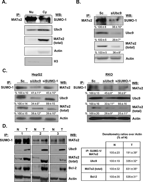
Figure 5. MATα2 is sumoylated in HepG2 and RKO cells and human colon cancers.

A. Nuclear and cytoplasmic proteins from HepG2 cells were immunoprecipitated with anti-MATα2 and Western blotting was carried out with anti-SUMO-1, MATα2 and Ubc9 antibodies. B. and C. HepG2 and RKO cells were treated with siUbc9 and siSUMO-1 or scrambled (Sc, 20 nM) for 48 hours and co-immunoprecipitation of MATα2 or SUMO-1 was performed followed by Western blotting with anti-MATα2 and anti-Ubc9 antibodies. Densitometric values are shown below the blots, and results represent mean ± SEM from 3 experiments done in duplicates expressed as % of Sc, *p < 0.04 vs. Sc. D. Sumoylation of MATα2 in human colon cancer specimens (T) and corresponding non–tumorous (N) tissues are shown. The immunoprecipitation was done as described above for sumoylated MATα2; total MATα2, Ubc9 and Bcl-2 are also measured in the same specimens using Western blotting. Densitometric changes are shown in adjacent box, and results represent mean ± SEM from 5 colorectal cancers expressed as % of corresponding non-tumorous (N) tissues, *p < 0.05 vs. non-tumorous tissue.
