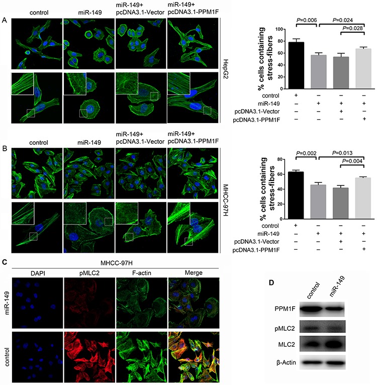
Figure 6. miR-149 regulated stress fiber formation, and PPM1F rescued the loss of stress fibers in HCC cells.

A, B. Immunofluorescent staining of stress fibers in HCC cells. MHCC-97H and HepG2 cells were transduced with miR-149 or negative control lentivirus, and transfection with pcDNA3.1-Vector or pcDNA3.1-PPM1F plasmid. Stress fibers were stained with Alexa Fluor 488-phalloidin, and cell nuclei were stained with DAPI. The percentage of stress fiber-containing cells was determined by counting 100 to 200 cells per experiment. Representative images (left panel) and data show the quantification of three independent experiments (right panel). C. The over-expression of miR-149 reduced the level of pMLC2 in MHCC-97H cells. Cells were transduced with miR-149 or negative control lentivirus, and cells were stained for F-actin with Alexa Fluor 488-phalloidin (green), pMLC2 with antibody (red), and nuclei with DAPI (blue). D. The levels of PPM1F, pMLC2 and total MLC2 were detected by Western blotting in MHCC-97H cells that over-expressed miR-149 or control cells. β-actin served as an internal control.
