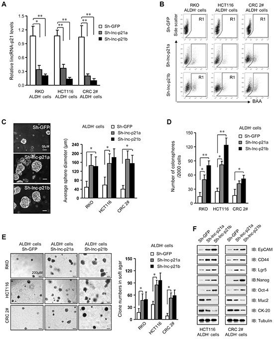
Figure 2. Knockdown of lincRNA-p21 enhances stemness and tumorigenicity of ALDH–CRC cells.

A. The level of lincRNA-p21 was evaluated by qPCR in ALDH− cells infected with lentivirus expressing two independent lincRNA shRNAs, Sh-lnc-p21a and Sh-lnc-p21b, at 10 MOI for 48 hrs. Infection with shRNA targeting GFP, Sh-GFP, served as control. B. Flow cytometric analysis of ALDH+ population in ALDH− cells after transduction for 7 days. C, D. The diameter (C) and numbers of spheres (D) generating by single ALDH− cells were measured with ImageJ software 14 days after infection with lentiviruses. More than 10 repeat wells were counted for each group and spheres with a diameter larger than 50 μm were included. E. Numbers of colonies formed by ALDH− cells with or without lincRNA-p21 knockdown in soft agar-containing medium. Colonies with a diameter higher than 75 μm were counted. F. Immunoblot analysis of stem cell markers and differentiation markers in ALDH− cells infected with lincRNA shRNAs or sh-GFP. Tubulin was a loading control. Representative graphs (B) or images (C, E, F) are shown. Data are presented as the mean ± SD (A, C, D, E) of each group from triple replicates. *P < 0.05, **P < 0.01.
