
Figure 6. Ad-lnc-p21-MRE suppresses glycolysis of CSCs through downregulating β-catenin/PDK1 signaling axis.
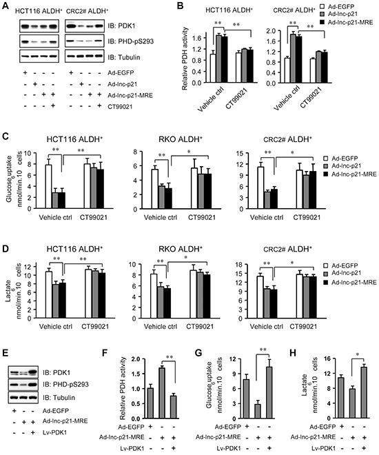
A. The levels of pyruvate dehydrogenase kinase 1 (PDK1) and phosphorylated PDH (S293) were detected by immunoblot analysis in ALDH+ CSCs, 48 hrs after treatment with Ad-EGFP, Ad-lnc-p21, or Ad-lnc-p21-MRE (10 MOI), or Ad-lincRNA-p21-MRE infection together with CT99021 incubation (3 μM). B. Relative PDH activity was determined in ALDH+ CSCs, 48 hrs after infection with Ad-EGFP, Ad-lincRNA-p21 and Ad-lincRNA-p21-MRE (10 MOI). CT99021 (3 μM) was simultaneously used to rescue the inhibition of β-catenin signaling by lincRNA-p21. C. ALDH+ CSCs were treated as in (B) and their glucose consumption was determined by a colorimetric assay kit. D. Lactate production by these ALDH+ cells was assessed with accutrend lactate analyzer. E. ALDH+ CSCs isolated from HCT116 cells were infected with Ad-EGFP or Ad-lnc-p21-MRE (10 MOI), or co-infected with Ad-lincRNA-p21-MRE and Lv-PDK1 for 48 hrs. The levels of PDK1 and phosphorylated PDH (S293) were detected by Western blot. F, G, H. ALDH+ CSCs isolated from HCT116 cells were treated as in (E). Their relative PDH activity (F), glucose uptake (G) and lactate production (H) were measured. Representative images (A, E) are shown. Data are presented as the mean ± SD (B, C, D, F, G, H) of each group from triple replicates. *P < 0.05, **P < 0.01.