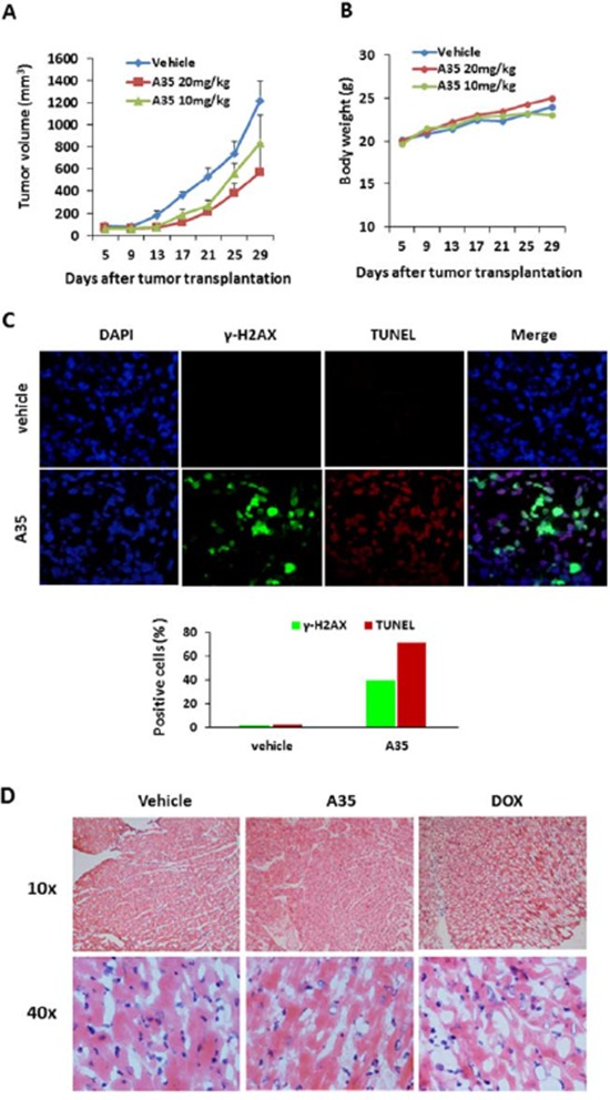
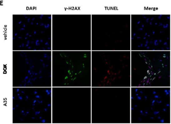
Figure 6. A35 suppresses tumor cell proliferation in vivo and has no toxicity in mouse hearts.


Nude mice (n = 5) bearing HepG2 xenografts were administered A35 on day 8 after tumor inoculation and successively administrated A35 for 21 days once per day. Mice were sacrificed when the tumor volume of the control group reached 1000 mm3; the tumor loads and hearts were isolated and prepared as frozen cardiac sections. A. Tumor volume was measured by calipers twice per week in the indicated days. B. Body weights of mice harboring tumors were monitored twice per week in the indicated days. C. γ-H2AX immunofluorescence and TUNEL staining of frozen tumor sections from various treatment groups; the total numbers of γ-H2AX-positive nuclei or TUNEL-positive nuclei as a percentage of the total number of nuclei are shown in the bar graph. n = 5 mice per group. D. H&E staining of frozen cardiac sections from various treatment groups. E. γ-H2AX immunofluorescence and TUNEL staining of frozen cardiac sections from various treatment groups.
