
Figure 3. Telomere shortening and dysfunction in HEL and BGC-823 cells treated with bortezomib, which was attenuated by hTERT over-expression.
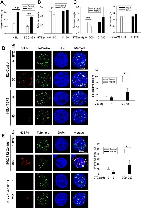
A. Enhanced telomerase activity in HEL and BGC-823 cells expressing ectopic hTERT. Cells were infected with lenti-viral or retro-viral hTERT-encoded vectors, and selected using puromycin. Telomerase activity was assessed using a telomerase PCR-ELISA kit. B. and C. Telomere shortening in bortezomib-treated control cells but not in hTERT-over-expressed cells. Cells were treated with bortezomib for 24 hours and FLOW-FISH and/or qPCR was performed to determine telomere length in these cells. B and C left panel: FLOW-FISH results; and C right panel: qPCR results. Telomere length in bortezomib-treated cells was expressed as percentages of that in untreated cells. D. and E. Telomere dysfunction in bortezomib-treated cells was inhibited by hTERT over-expression. Control and hTERT-over-expressed cells were treated with bortezomib for 24 hours and then analyzed for co-localization of 53BP1 foci and telomere signals (TIF) using immuno-FISH. Cells were counted for TIF and the percentage of positive cells (> 2 foci/cell) calculated (right panel). Shown in (D) and (E) left panels is representative of three independent experiments. * and **: P < 0.05 and 0.01, respectively. BTZ, bortezomib.