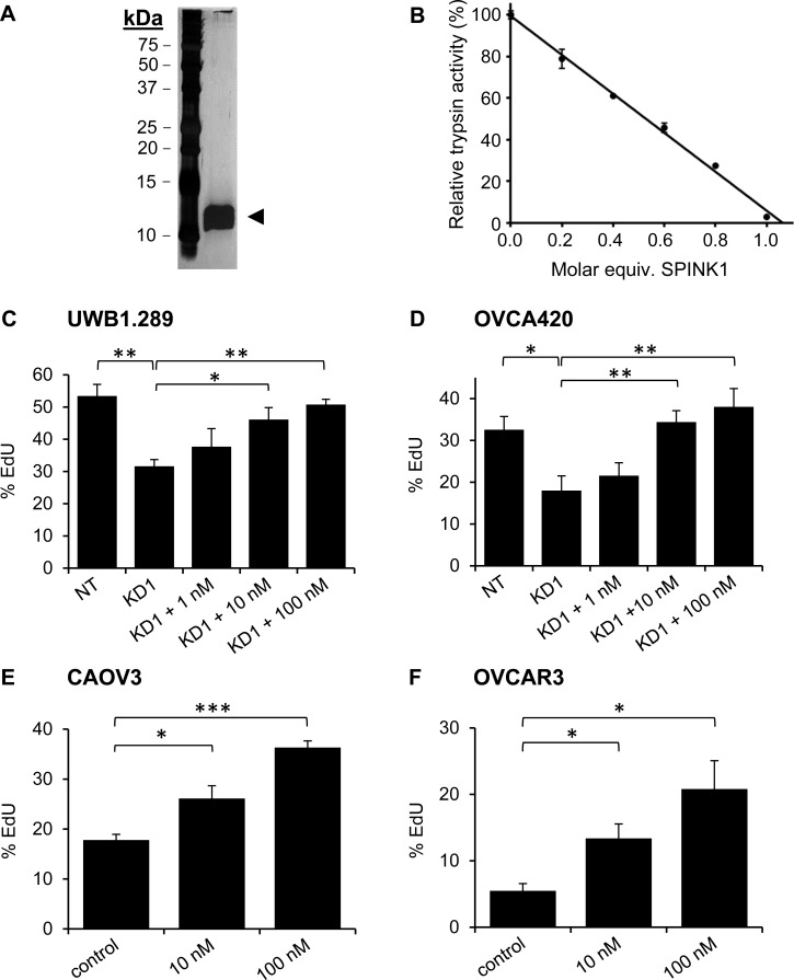
Figure 2. Recombinant SPINK1 stimulates proliferation of ovarian cancer cells.
A. Silver stained gel shows highly purified rSPINK1 protein following expression in HEK293E cells and purification via affinity chromatography and gel filtration. B. rSPINK1 shows full inhibitory capability against bovine trypsin at equimolar concentration. C., D. Reduced proliferation in UWB1.289 cells C. and OVCA420 cells D. after transduction with SPINK1-targeted shRNA (KD1) is rescued by treatment with rSPINK1 at concentrations indicated, as assessed by EdU assays. UWB1.289 NT vs KD1 p = 0.0062; KD1 vs 10 nM p = 0.0271; KD1 vs 100 nM p = 0.0017. For OVCA420, NT vs KD1 p = 0.037; KD1 vs 10 nM p = 0.013; KD1 vs 100 nM p = 0.0108. E., F. CAOV3 and OVCAR3 cells treated with different concentrations of rSPINK1 show dose dependent increases in proliferation as assessed by EdU assays. (CAOV3: 10 nM, p = 0.0246, 100 nM p < 0.0001, OVCAR3: 10 nM p = 0.0183, 100 nM p = 0.0137.) *p < 0.05; **p < 0.01; ***p < 0.0001 (unpaired t-test).

