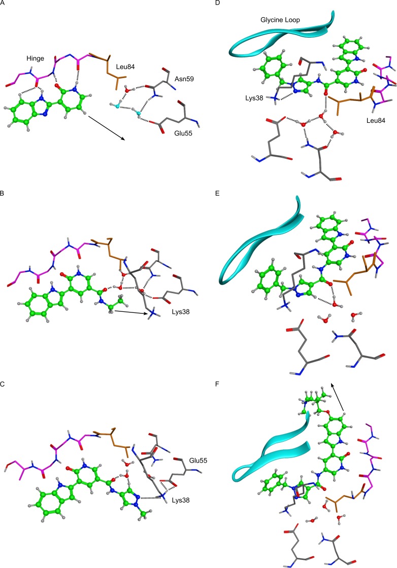
Figure 2. X-ray crystal structures of key molecules in evolution of VER-154637 to V158411.
Hydrogen atoms were added to the X-ray coordinates with the software MOE, and only selected hydrogens are shown. Dotted lines indicate inferred hydrogen-bond interactions, and arrows indicate vectors used for structure-guided chemical elaboration. Key amino acids and structural features are indicated. In panel A, the two water molecules with light blue oxygens were modelled by analogy with the three conserved water molecules observed in most Chk1 X-ray structures. A. VER-154637. B. VER-154931. C. VER-155175. D. VER-155422. E. VER-155991. F. V158411 (PDB ID: 5DLS).

