
Figure 7. Recellularization of kidney scaffolds with mouse ES cells.
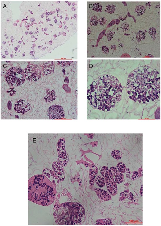
Hematoxylin and eosin staining of kidney scaffold seeded with mouse ES cells show homogeneous distribution of cells into glomerular, vascular structures, peritubular capillaries, and tubules. A. shows the gross appearance. B–D. shows the glomeruli part and E. shows the renal tubule.