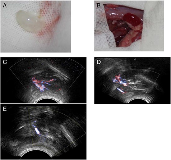
Figure 8. Implantation and explantation of the renal ECM scaffolds.

The gross appearance of the scaffold A&B. and the blood flow shown by B-scan ultrasonography C–E. (A), The acellular scaffold prior to implantation. (B), After removal of the clamps, blood flowed well within the whole scaffold. B-scan ultrasonography images of the implanted kidney scaffold obtained on day 2, weeks 1 and 2.
