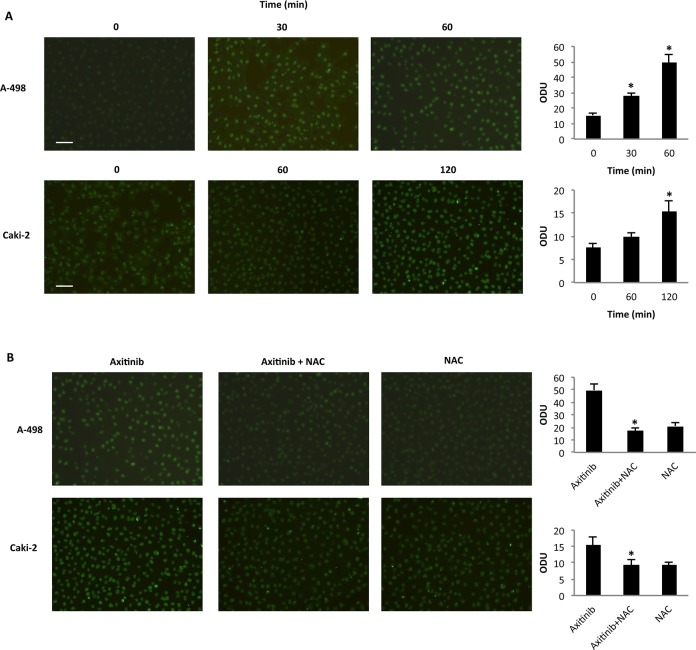
Figure 3. Axitinib triggers oxidative DNA damage in RCC cells.
A. Immunofluorescence analysis of 8-oxo-dG in A-498 and Caki-2 RCC cells treated with axitinib (12.5 μM and 25 μM, respectively) for different times. Optical Density Units (ODU) were calculated on ten random fields. Data shown are representative of one of three separate experiments, *p < 0.01 vs. untreated cells. Bar: 100 μM. B. Immunofluorescence analysis of 8-oxo-dG in A-498 cells treated with axitinib 12.5 μM for 60 min and in Caki-2 cells treated with axitinib 25 μM for 120 min alone or pretreated with NAC (10 mM for 1 h). ODU were calculated on ten random fields. Data shown are representative of one of three separate experiments, *p < 0.01 vs. axitinib treated cells. Bar: 100 μM.

