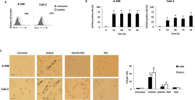
Figure 5. Axitinib induces cellular senescence in RCC cells in a ROS-dependent manner.
A. Representative flow cytometric profiles of RCC cells untreated or treated with axitinib for 72 h, then stained with C12FDG, a fluorogenic substrate for SA-β-galactosidase before analysis. Data represent the percentage of positive cells. B. ROS generation in RCC cell lines treated with axitinib for the indicated times. Cells were stained with DCFDA before flow cytometric analysis. Data are expressed as percentage of DCFDA positive cells with respect to untreated cells; *p < 0.01 treated vs. untreated cells. C. Cellular senescence was assessed in A-498 cells treated with axitinib 12.5 μM and in Caki-2 cells treated with axitinib 25 μM for 72 h, with or without pretreatment with NAC 10 mM for 1 h, by detection of SA-β-galactosidase activity using cytochemistry. Arrows indicate blue-stained positive cells. Graph represents the percentage of β-galactosidase+ cells calculated on ten random fields. Data shown are representative of one of three separate experiments, *p < 0.01 vs. untreated cells. #p < 0.01 vs. axitinib-treated cells. Bar: 25 μM.

