
Figure 3. GIT1 regulates tumor growth and metastasis in orthotopic animal models.
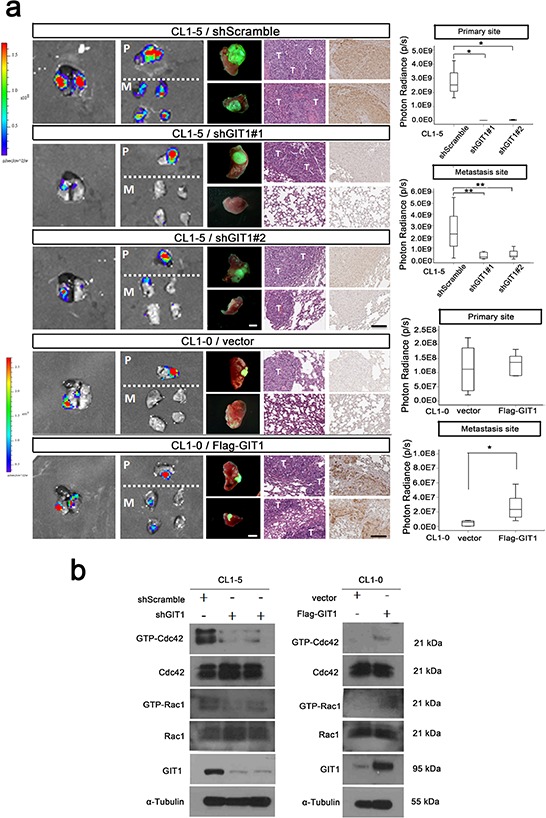
We generated the CL1–5-GL and CL1–0-GL cell, which were stably expressing GFP and luciferase proteins and could detect the GFP and Luciferase signal simultaneously. We performed in vivo orthotopic model experiments by injecting CL1–5 and CL1–0 cells (1 × 10⁶) with or without GIT1 knockdown or expression into NOD/Shi-scid/IL-2Rγnull (NSG) mice at the left side lung and determined the metastasis to contralateral lung. a. Establish GIT1 knockdown CL1–5/shGIT1 and GIT1 overexpression CL1–0/Flag-GIT1 cells as described in Figure 2b. shScramble is CL1–5 group control; Vector is CL1–0 group as control. Representative photon images of lungs were taken 4–6 weeks after orthotopic injection of the indicated CL1–5 and CL1–0 cells into NSG mice. Mice were subjected to luciferase imaging (Left). GFP signaling (Middle), Representative mice lungs and H&E and GIT1 IHC staining were shown in each group (Right). Scale bar: 100 μm. The photon signals of lung metastases were quantified in each group (Primary and metastasis site). *P < 0.01. **P < 0.001; n = 8 mice per group. Abbreviations: P, primary site; M, Metastasis Site; T: Tumor site. b. In vivo orthotopic mice Rac1/Cdc42 activity assay.